The measure of a digital pathology scanner’s colour reproducibility.
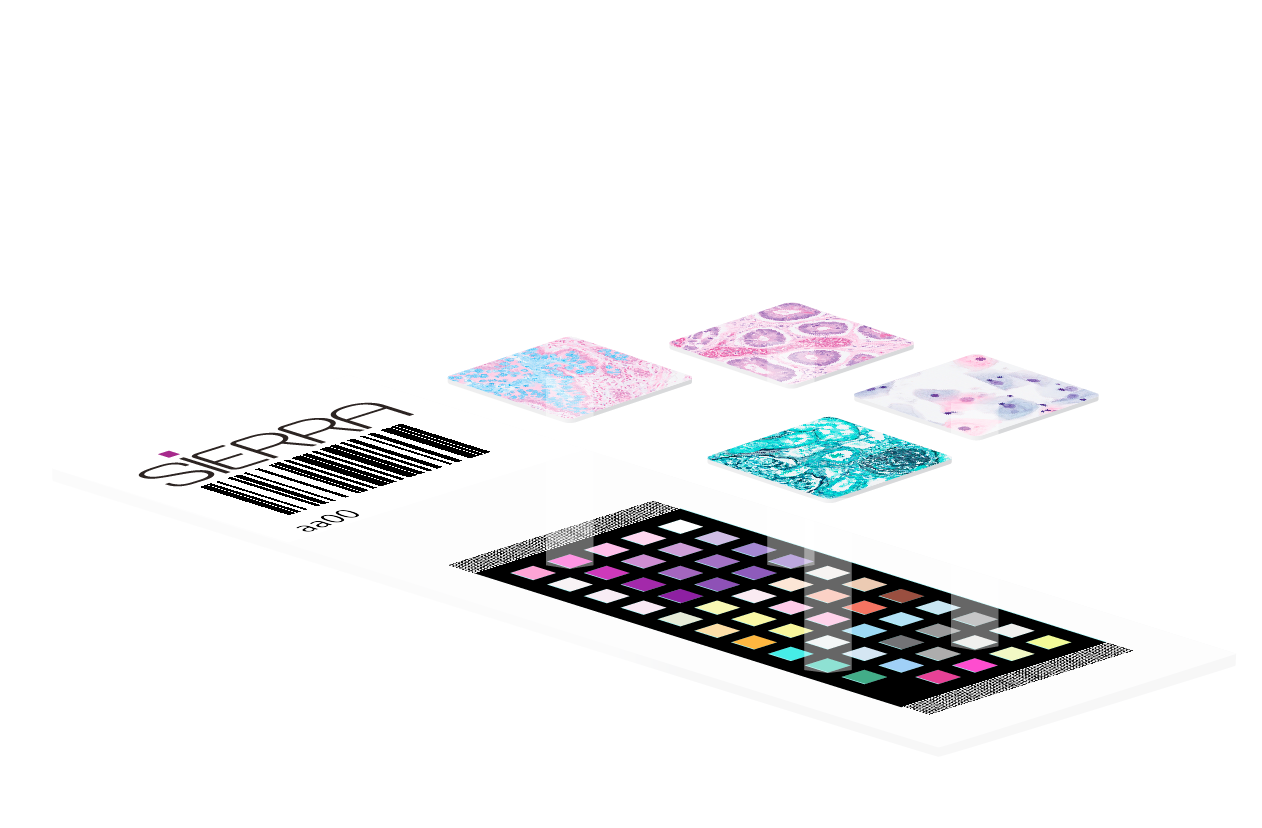
The Sierra slide carries 55 biopolymer patches, which mimic the spectral response of human tissue when stained for histopathology.
The spectral absorption of commonly used pathology stains is then measured using a traceable calibration spectrophotometer. This process captures the ‘ground-truth’ colour of the different stained patches, which can then be compared to those reproduced by a scanner.
Formatted like a pathology slide, so compatible with Whole Slide Imaging (WSI) Scanners:
Sierra Slide
Formatted like a pathology slide, so compatible with Whole Slide Imaging (WSI) Scanners
Small patches of biopolymer bind pathology stains and mimic the spectral response of stained tissue.
Creates gamut of pathology colours with reduced metamerism.

Stained with same protocol as pathology.
Learn about our other products
Sierra VS-Analyser
Sierra VS-Analyser validates colour of whole slide imaging devices and reports the difference between image colour and ground-truth colour in different scanners over time providing traceable Quality Assurance.
Sierra VS-Profiler
Sierra VS-Profiler generates an individual ICC profile for each whole slide imaging device the Sierra Slide is scanned on, standardising the colour of the digital images produced by any scanner to ground-truth colour.
Contact Us
For more information on all of our Sierra Slide products or any other questions please contact us using the following form